
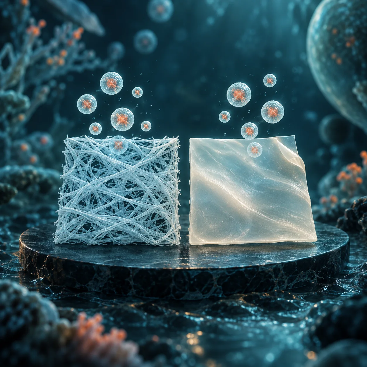
How does carrier structure affect the release of extracellular vesicles (EVs)? A study published in Frontiers in Bioengineering and Biotechnology on July 7, 2026 loaded EVs into dry silk-fibroin nonwoven materials and films, comparing release over 14 days and in vitro cellular responses.
Two structures, two release curves
The researchers used EVs derived from gingival fibroblasts and treated the silk-fibroin carriers with mild water-vapor annealing. The nonwoven material showed relatively gradual, controlled release. The films showed an initial delay followed by faster release. Released EVs retained their morphology and could be taken up by the human umbilical vein endothelial cells used in the experiments. Original study
In vitro scratch assays also found that both EV-loaded materials promoted cell migration. This measures cell behavior under culture conditions and cannot substitute for animal or human wound outcomes.
Material design must consider the full period of use
Even when both carriers contain EVs, different structures may produce different release timelines. Local-delivery materials therefore require consideration of loading, release onset, release rate and the state after release. One time point cannot capture the whole process.
TASCAP considers this work a concrete approach to comparing carrier structure with release behavior. If the EV source, drying process or final formulation changes, the new combination will need its own evaluation.
The subjects of this study were gingival-fibroblast-derived EVs and silk-fibroin carriers. Sharing a dry-storage characteristic does not make its 14-day release results automatically applicable to other freeze-dried products. The conclusions remain specific to this material and process combination.